Till date, a lot of impenetrable mysteries still surround the nature of the cherry eye ailment. The anatomical analysis of the ailment shows that it’s related to a defective nictitating membrane. The nictitating membrane (also known as the third eyelid), is a connective tissue underlining the lower eyelids of dogs. It is responsible for eye lubrication and the production of the tear film, and also acts as a protective shield against dust and other foreign objects. The third eyelid also comes with a tear gland that supplies the dog’s eye with 35%-50% of the total moisture required. Hence it’s a crucial component of the dog’s ocular system.


Cherry eye in dogs
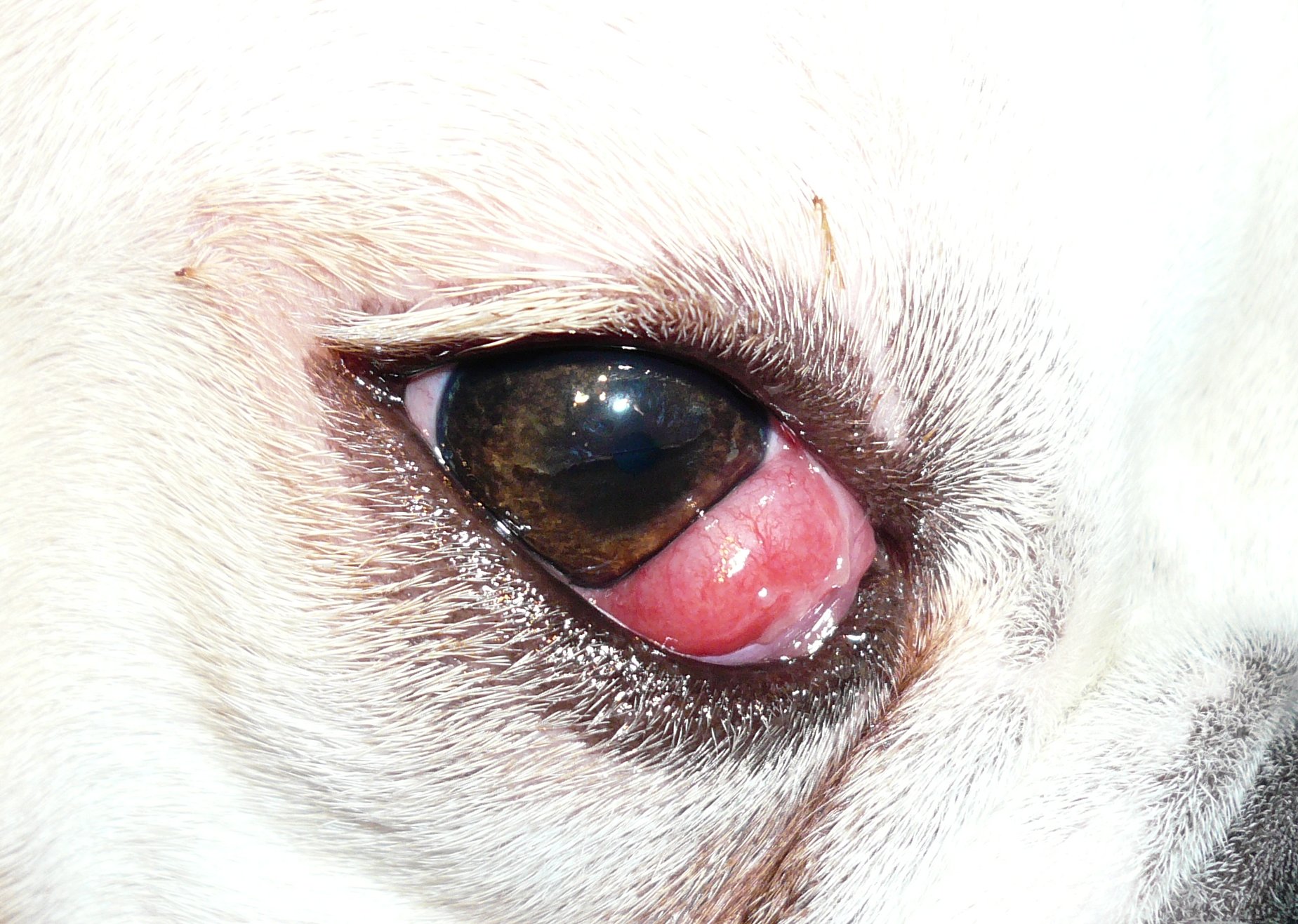 But when the bond between the fibrous tissues and the eye-globes weaken, the tear glands protrude through the pockets of spaces between the two disentangling parts. This severance allows red tissue masses to spurt across the visible eye sections. The partial eclipse usually covers the visible eye section nearest to the nose. Also, as a natural response to such breakdown of links between connective tissues, tissue hypertrophy ensues, as the nictitating membrane swells, not because of a proliferation of cells, but the bloating of extant cells. This outlay can spread across both eyes and leaves the affected dogs desperately scratching and pawing away at the affected site. The ailment’s moniker does a very quirky job of capturing the creepy bulbous red pigment that characterizes the ailment.
From all indications, cherry eyes seem to belong to genetic pathology. Its incidence is more frequent in certain species, even though every dog is susceptible to it simply because its caused by the collapse of tissues.
Safeguards Against Cherry Eyes
There is no precise knowledge about the processes that bring about the collapse of the nictitating membrane. But an outstanding fact about the ailment is that it is frequently observed in certain breeds.
The symptoms of the ailment, including eye redness, inflammation, and hypertrophy, are all minor health conditions. However, a dog should be given proper medical attention should it exhibit symptoms of the ailment. If the case recurs in a dog, the symptoms can worsen over time. An untreated cherry eye can lead up to more critical ailments in the long-run. If the third eyelid is exposed in a protruding pose for too long, there’s likely going to be a decrease in moisture production, and that can culminate in serious ails.
Also, the protruding membranes restrict proper blood circulation in the affected area, and this can bring about more serious complications if left to continue. It can also cause the onset of secondary bacterial infections.
If you’re able to spot the symptoms of the ailment early enough, you can nip it in the bud. Simply massage the protruding tear gland with a warm, moist cloth and dog eye drops, until the misplaced nictitating membrane snaps back into place. However, this simple home remedy is a short-lived solution, and the risk of recurrence lurks around. The appearance of symptoms of the ailment call for an urgent medical response.

Conclusion
The symptoms of cherry eyes usually emerge abruptly. From one minute to the next, a surge of seething red tissues might be seen launching out of the bottom or corner of the eyelid of a dog who’s been healthy and bouncing all along.
However, the genetically induced cherry eye can come in the form of a birth defect in which the third eyelids are visibly seen in the lower edges of a newly born dog’s eyes. The sight is commonly labeled “haws.” This, however, cannot be properly termed a cherry eye, since it has only aesthetic and cosmetic consequences. Dogs with haws tend to appear lethargic, lackluster and dispirited, and thus they’re generally unattractive.
The causes of cherry eyes are poorly understood. But it's a well-documented fact that cherry eyes can take a hereditary dimension and pass on from one generation to the next. It tends to occur frequently in breeds with compact muzzles. But all dogs at all life stages are susceptible to the ailment. Beyond the genetic factor and the anatomical analysis of the ailment, nothing much is known about its true nature. It's certain that failure of the connective tissues that enclose the nictitating membranes leads to a protrusion of the tear gland, and this condition most likely holds in breeds with certain genetic makeups.